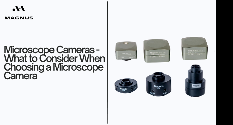
Microscope Cameras - What to Consider When Choosing a Microscope Camera By Magnus Opto

In recent years, medical and life science fields have faced significant demands to accurately capture and process microscopic images. So, what's essential? It begins with selecting the appropriate camera.
Choosing the right microscope camera can be a daunting task, especially with the numerous options available in the market. The selection process requires careful consideration of several factors to ensure that the chosen camera meets the specific needs of the application.
Here you will explore the key considerations for selecting the ideal microscope camera for your specific needs.
What is a microscope camera?
A microscope camera is a specialized device crafted for use with microscopes. It aimed at capturing high-resolution images or videos of microscopic specimens for analysis, study, and sharing purposes.
Available in various forms, such as digital cameras attachable to a microscope's eyepiece or integrated directly into the microscope, they offer flexibility in capturing and documenting microscopic observations.
Additionally, these cameras often include accompanying software for advanced image processing and analysis. It enhances their utility in research, education, and clinical settings.
Sensor Type and Size
The most suitable sensor type and size provide a higher level of detail in the capture of living cells and tissues. This enables more accurate analysis of biological processes. Today, CCD and CMOS are the two main types of image sensors used in cameras.
CCD stands for Charged Coupled Device. In CCD sensors, a charge accumulates in each pixel during image capture, and the data is read sequentially once the capture is complete. CMOS stands for Complementary Metal-Oxide Semiconductor.
CMOS sensors read charges simultaneously using a parallel approach. CCD cameras excel in light sensitivity and noise reduction. However, CMOS cameras deliver images faster due to their parallel processing and are typically more affordable, which is ideal for simpler digital imaging needs.
High Resolution
Camera resolution defines the level of detail an image can provide. When using a microscope to capture detailed images of cell structures, tissues, or organisms, it is essential to match the camera's resolution with the microscope's resolution.
The pixel size required by the camera depends significantly on the resolution and magnification of the microscope objective. If the camera resolution is excessively high, you waste pixels and storage space.
Conversely, if the camera resolution is too low, it fails to capture sufficient detail at lower magnifications. Therefore, selecting a camera that aligns with the desired resolution is crucial to achieving optimal image quality.
Camera Noise
The next thing you need to consider when buying microscope camera is camera noise. Camera noise is classified into three types. They are dark noise, read noise, and shot noise.
In fluorescence microscopy, microscope cameras with low read and dark current noise are highly sought after because they produce clearer images.
This can take advantage of a higher gain. Lower read noise allows for higher gain usage without degrading image quality. It leads to more precise and reliable imaging results.
Pixel Size
Pixel size represents the dimensions of individual photodetectors. It is a crucial consideration in camera selection. It directly impacts the spatial resolution limit of the sensor. Smaller pixel sizes result in finer image details at any given magnification.
However, decreasing pixel size reduces the sensor's photon capacity. It limits the number of photons it can detect without saturation. Consequently, smaller pixels often exhibit lower dynamic range and poorer signal-to-noise ratios.
For optimal performance, a high-resolution camera with a pixel size around 6.5 µm typically offers sufficient resolution. This balance ensures detailed imaging without compromising on image quality due to noise or a limited dynamic range.
Color Reproduction
Camera sensors have a spectral response that differs from that of the human eye. It requires camera manufacturers to use a variety of ways for correct color representation on computer screens, similar to what is seen under a microscope.
Key features include automated white balance, which simplifies operation by detecting and correcting bright backgrounds without the need for operator intervention. Multi-axis color adjustment allows for exact color optimization for various stains.
It distinguishes between hues such as red and brown for a separate presentation inside the image. Furthermore, color spacing and matching functionalities broaden the camera's color range, which is essential for vivid stains and ensures the correct portrayal of a wide spectrum of colors.
High Sensitivity
In fluorescence microscopy, high sensitivity in a microscope camera is essential to prevent sample photobleaching and capture high-quality images even in low-light conditions. Sensitivity is determined by quantum efficiency.
This efficiency measures the sensor's ability to convert photons into electrons. A larger light-sensitive area on the sensor leads to higher QE and increased sensitivity. It is essential to check the recent sensor advancements. It further enhance sensitivity.
These improvements allow for more efficient detection of faint signals. It results in clearer and more detailed images of biological samples in fluorescence microscopy applications.
Quantum efficiency
Quantum efficiency characterizes a camera's ability to convert incoming light photons into electrical charge. Achieving a QE of 100% would mean perfect conversion of every photon to an electron.
However, QE varies across the spectrum, with CMOS and CCD detectors exhibiting lower efficiency towards the wavelength range's extremes. The maximum QE typically observed in cameras is approximately 0.6.
This limitation underscores the importance of considering QE when selecting a camera. Despite this limitation, modern camera technology strives to optimize QE to enhance overall performance and sensitivity in capturing and detecting light signals across the spectrum.
Compatibility and Connectivity
Compatibility and connectivity are crucial considerations when selecting a microscope camera. The camera must be compatible with your microscope model, which may involve specific mounting options or requirements.
The connection type is also vital, as it determines how the camera interfaces with your computer or microscope. USB is a widely supported and straightforward option, while HDMI may offer higher data transfer rates.
Ensure that the camera is compatible with your microscope model and choose a connection type that aligns with your needs, which ensures seamless integration and optimal performance.
Final Words
Selecting the right microscope camera requires a careful balance of factors. By considering above mentioned key aspects and aligning them with your specific imaging needs, you can choose a camera that satisfy your needs. With the right microscope camera, you can unlock the full potential of your microscope and gain valuable insights into the microscopic world.


